Wyszukaj w wideo
“My unusual” patient with heart hypertrophy
II Międzynarodowy Kongres Kardiomiopatii – warsztaty z rezonansu magnetycznego serca
Podczas II Międzynarodowego Kongresu Kardiomiopatii eksperci omówili najważniejsze zagadnienia dotyczące tych schorzeń. Jakie trudności niesie ze sobą opieka nad pacjentem z kardiomiopatią?
Odcinek 4
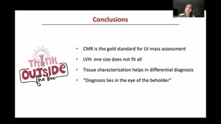
Dr Anna Baritussio (MD, PhD) przedstawiła nietypowy przypadek pacjenta z przerostem serca, podkreślając znaczenie zaawansowanej diagnostyki obrazowej. Omówiła kluczowe ustalenia z badania CMR, w tym asymetryczny przerost lewej komory (LVH), obecność ogniskowego LGE w przegrodzie oraz podwyższone wartości T1 i ECV. Wykład zwracał uwagę, że CMR jest złotym standardem w ocenie masy lewej komory i różnicowaniu kardiomiopatii. Dr Baritussio podkreśliła, że w diagnostyce "one size does not fit all", a charakterystyka tkankowa odgrywa kluczową rolę w rozpoznaniu. Wnioskiem było również, że trafna diagnoza wymaga doświadczenia i indywidualnego podejścia do pacjenta – "Diagnosis lies in the eye of the beholder".
Good evening, everyone.Uh, thank you very muchfor the opportunity to share thisunusual case.
This is a case ofa 38-year-old male.Uh, his past medical history,um, was, um, remarkable for type1 neurofibromatosis and for T-cellacute lymphoblastic leukemia, for which thepatient was in his firstyear of remission.
He presented because of diplopiaand hand vasculitis, and because ofthis, he received a brainMR that showed edema of rectusmuscle of the eye.
This is his ECG onpresentation.As you see, we havewidespread T-wave inversion, not only inthe, uh, precordial leads butalso in the inferior leads.
Given the hand vasculitis anddiplopia, a transthoracic echo was requestedto rule out sources ofemboli.
Uh, the echo did notshow valvular vegetations, but it showedthe presence of biventricular hypertrophywith some speckled, uh, appearing myocardium.So, a CMR was requestedto rule out cardiomyopathy.
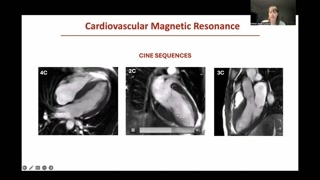
So, we start to havea look at CMR, uh, imagesfrom cine sequences.You can see here, thelong axis, four, two, and threechamber.What we appreciate here isthat eyeballing normal biventricular function withvery much a- asymmetric hypertrophyof the left ventricle, involving notonly the septum, but also,as you see here, the basalto mid-inferior wall.If we look here atthe valves, tricuspid, mitral, and aortic,we agree with echo thatwe don't see, uh, much, um,valvular abnormalities.
Uh, biventricular function looks normal,but you do know that weneed to confirm, uh, uh,this also on the short axiscine.You see here from baseto the apex, that we havenormal biventricular function, EF ofthe left ventricle of 68%.We confirm the presence ofasymmetric left ventricular hypertrophy, mainly involvingthe septum, and of theseptum, mainly involvement- involving the inferiorseptum towards or extending towardsthe, uh, inferior wall.And you see here thatwe got a maximal wall thicknessof 21 millimeters here inthe, um, inferior septum.
Of course, you do knowthat the key point of CMRis tissue characterization.
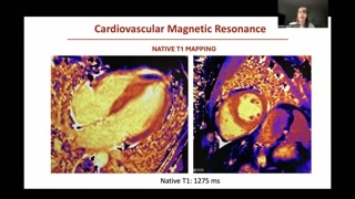
Uh, we can nowadays performparametric mapping.Parametric mapping can have differentweighting, and this is a nativeT1, meaning by that, thatwe provide these images before givingcontrast, which means that wecan get information also without administeringcontrast, and this is particularlyimportant for those patients that cannotreceive, um, contrast agents, forexample, because of impaired, uh, renalfunction.
This native T1 map createsa color-coded map that gives usa visual idea of whetherthe myocardium has normal or abnormaltissue characterization, and for yourreference, normal myocardium should have thispurple appearance, whereas this orange,and the higher- the lighter theorange, the worse the case,uh, the- the orange signal meansthat we have an increasednative T1 value.
Increased native T1 value isabnormal, because it means that theinterstitium is expanded, and weusually find, uh, increased native T1values in cases such ashypertrophic cardiomyopathy, amyloidosis, whereas we findvery low native T1 mappingvalues in patients, for example, withFabry disease.So, we can measure sometimes here in milliseconds, and inthe case of the patient,the native T1 mapping is quiteincreased, because it's above 1200.
We're probably more familiar withthe post-contrast sequences, where we havethe, um, the, um, uh,cavity that looks, uh, brighter becausethere's a contrast in it.The myocardium appears black ordark, uh, gray, and if wehave late gadolinium enhancement withinthe myocardium, we have these brightsignals, as spotted here bythis arrow, that shows a mid-wall,therefore non-ischemic LGE of thebasal to mid-cavity inferior septum.
But, what really, uh, catchesthe eye are all these nodularareas that affect, quite extensively,the left ventricle, that are really,really very dark.They're darker than the myocardium,so more hypointense than the myocardium,and they involve not onlythe left ventricle, but also theright ventricle and the epicardium.
As I told you, wecan get different weighting with these,uh, with these mappings, butwhat we may do is thatwe acquire the nat- theT1 mapping pre- and post-contrast.And why do we doso?Well, first of all becausewe get an idea that somethingvery odd is going onin this left ventricle, but alsowe c- because we canget, um, an estimation of theextracellular volume, so we canmeasure this expansion of the interstitialspace.And in the case ofour patient, this is, uh, 40%,so it's increased in keepingwith the increased native T1 values.
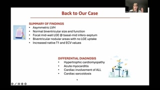
So, to summarize the findingsof CMR, we have an asymmetricleft ventricular hypertrophy.We have normal biventricular sizeand function.
We have focal mid-wall, thereforenon-ischemic LGE of the basal andmid-inferior septum, and we havethis, um-...Numerous areas, nodular areas thatdo not pick up contrast alongwith increased native T1 andextracellular volume values.
Does this point to asingle cardiomyopathy?Probably hard to say atthis stage but we can getup to a differential diagnosis.
So this could be acase of hypertrophic cardiomyopathy.Why not?This could be a caseof chronic sarcoidosis, LVH possible, acutemyocarditis, could be, or maybethis could be just an involvementof the hematologic disease ofthe patient.
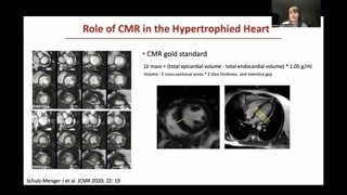
So when it comes to,uh, an hypertrophic heart, CMR isvery useful.First of all because alongwith being the gold standard forvolume and function assessment, CMRis a gold standard also forthe assessment of LV mass.And this is because itis a technique that is freeof any geometric assumption, andwe basically contour the epicardium andendocardium of the left ventriclein systole and in diastole.We do it, uh, foreach slice to cover the entirevolume of the, um, ofthe left ventricle, and then withthis formula we get theLV mass.
But another important thing isthat sometimes there are areas ofthe left ventricle that wecannot explore as easily with echo.Say, for example, the infraseptumwhich is the most involved, uh,on CMR in this patient.Sometimes we miss the hypertrophyon echo while this is clearlyseen on CMR as you'veseen in this case.
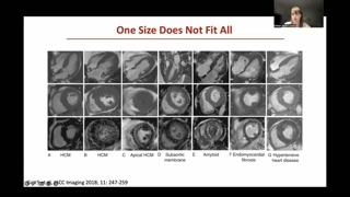
But the other point isthat sometimes just based on echoor based also on thecine sequences in CMR, um, thehypertrophied heart may have verysimilar appearance- appearance.Take, for example, the thirdand the sixth case, but itis only by providing myocardialtissue characterization that we find thatall these parts do referto very different cardiomyopathies.So if we took theexample of the third and sixthcase, in this case, wehave an apical hypertrophic cardiomyopathy andthis is an endomyocardial fibrosis.Very different diseases that maylook very much similar on echobut also on cine CMR.
And so it is reallytissue characterization that makes a hugedifference because we can basicallyassess all tissue abnormalities, although webasically look for two thingswhich is myocardial edema by usingthe T2-weighted and the T2mapping sequences, and then we lookfor myocardial fibrosis or necrosisor injury according to diseases bylooking at T1 mapping, lateenhancement, and deriving the extracellular volumeby looking at the nativepre- and the native and thepost-contrast T1 mapping.
So back to our casewe said asymmetrical VH, normal sizeand function.We have some non-ischemic LGbut it's very minimal, focal whilewe have these many nodularareas that are looking very oddwithout picking up contrast.So these are our differential.Let's walk through each ofthose to see whether we can,um, manage to understand what'sgoing on.
So acute myocarditis well couldbe an option.We have abnormal ECG.The patient didn't actually havechest pain but you know sometimesthe patient presents with embolism.Uh, it comes from amyocarditis so we have to havea very open mind butwe do know that CMR offerssome specific criteria.We have the updated LakeReese criteria where we need oneT2 criteria so evidence ofmyocardial edema whether it is onT2 standard T2 weighting orT2 mapping and we need, uh,one criteria for non-ischemic myocardialinjury so one T1 criteria thatcould be increased native T1or, uh, um, positive LGEs witha non-ischemic pattern or abnormalECV.To be honest with youour patient definitely has some EC-some LGE, has the increasednative T1, has the increased ECV.Unfortunately we did not perform,um, the T2 weighted images sowe don't know whether thereis or not edema so wecan't really rule out myocarditisin this patient but you mightagree with me that there'ssomething that looks really, really odd.
One thing that is importantto bear in mind when itcomes to myocarditis is thatsometimes you see myocarditis patients withload of hypertrophy of theleft ventricle in the acute phaseand this is due tomyocardial edema.So this is an exampleof a case I'm sorry thisdoesn't play but this isan acute presentation heart failure yousee how hypertrophied uh theuh left ventricle looks like andthis is um a T2mapping for myocardial edema where normalmyocardial should be orange buthere it's all yellow because T2values are really really increasedshowing extensive myocardial edema and thispatient had a lymphocytic ummyocarditis that was properly treated withimmune suppression and you seehere the one week follow-up oneweek follow-up where we havea complete resolution of LVH becauseyou see here on thatstandard T2 weighted that we don'thave any myocardial edema leftso remember that LVH might indicateedema in the acute phasesof myocarditis but I will putit at aside on theother differentials we also had hypertrophic
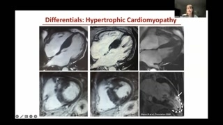
cardiomyopathy.We do know that wehave specific criteria, specific cut-off pointsfor hypertrophic cardiomyopathy above 15millimeters our patient had 21.We can have basically areally a wide way of presentingLVH because it can besymmetric asymmetric focal localized just towardsthe apex but typically hypertrophiccardiomyopathy has chunky non-ischemic usually mid-wallor epicardial but usually mid-walllead enhancement of the hypertrophied areasas you see here inthese cases while usually...The apical form may havesome transmural late enhancement of theapex.It's specifically, uh, if thereis an LV, uh, apical aneurysmas is in this case,where we usually spot the presenceof LV thrombus.
Um, last but not least,uh, we may have also cardiacsarcoidosis, right?So here there is, um,this is a still image froma cine sequence.You see here that thereis something that resembles our case,because we have an apertrophic-an hypertrophic left ventricle, but thisis an asymmetric hypertrophy.You see here that itinvolves mainly the septum and specificallythe inferior septum, and thiscorresponds to a wide area oflate enhancement with a non-ischemicsubepicardial pattern of the, um, the,uh, septum with a typicalpatchy appearance on the, uh, longaxis.So again, this may resembleour case because of the asymmetrichypertrophy, but the late enhancementdoesn't really ring a bell, uh,compared to the one ofour patient.
Myocarditis, I'm not really verymuch convinced.Hypertrophic cardiomyopathy, yeah, buta very weird one.Uh, sometimes, you know, ifwe're not sure because, you know,the cine sequence of ourpatient could be very similar toa- this sarcoidosis case, butwe can use other imaging modalitiesas, for example, PET, PETscan, that may show active, um,sarcoidosis with increased, uh, SUF,uh, S- uh, SIF values.Uh, but on the otherhand, we may use CMR alsoto look for external, um,findings and features that may supportthe suspicion of cardiac sarcoidosisas, for example, the presence oflymphadenopathy, uh, in the thorax.
But we need to goback to our case, because weneed to put pieces together.
So we said we haveasymmetric hypertrophy.There are these nodular areason, uh, uh, diffused, uh, inthe myocardium, and these areaslook iso-intense to the myocardium onthe cine sequences, iso-intense tothe myocardium on these localizer thatare mainly T1 weighted sequences,and they do not pick up,uh, they do not pickup contrast on late enhancement sequences,so they're very, very, uh,very dark.
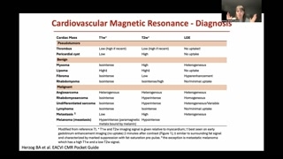
So what could this be?Well, if we think ofmasses, because these are nodular areasand we think of cardiacmasses, uh, could be benign, couldbe malignant, there are nicetables to try and work outwhat masses may be.Remember that CMR does notprovide histology, but the tissue characterizationcan go down to acouple of possible differential diagnoses tohelp also the clinicians tochoose the proper...For the test.
And if we go throughthis list, what we see hereis that sometimes hematological, um,tumors, lymphomas may have a behaviorthat's very similar to thisone with a iso-intense presentation onthe T1 weighted images andwithout, or very minimal uptake onthe late enhancement.So we have a patientthat has a history of leukemia.He was treated.He was in his remissionphase.
But let's think about it.So what we did we,we went through the scan, becausewe don't just look atthe heart, right?We l- we don't limitourselves.We don't put our headinto boxes.And what we found, uh,is that, uh, there was aleft kidney enlargement.Why?Very weird.And we remember that thepatient came in with diplopia andhe had edema of therectus muscle.
So we thought what ifthe patient has a relapse ofhis hematologic disease?So can we have anyhistological proof of, uh, of thedisease?Is there anything we canbiopsy?And what we did isthat we get the biopsy ofthe muscle, and this confirmedthe presence of acute leukemia.So we concluded that thiswas a case of a multi-organrelapse of T-cell acute lymphoblasticleukemia.
So the patient was treatedwith steroids.He started systemic nilarabine andalso had treatment, uh, intrathecally.
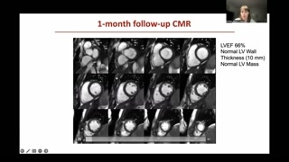
He had a rapid improvementclinically, but also on imaging.Brain imaging showed complete resolutionof the extraocular muscle enlargement, sowe repeated CMR one monthlater, and this is the picture.
So you see here, wedon't have any more the asymmetrichypertrophy of the left ventricle.You see that it lookspretty much more homogeneous.The function is still preservedby ventricular function.We check also on theshort axis because we're always checkingtwo orthogonal planes, and yousee here that we confirm thatthe thickness is back tonormal, 21 to 10 millimeters.Normal LV mass was increaseda big- at the beginning, butluckily enough, irrespective of chemotherapy,LV function and RV function aswell, still preserved.
But let's have a lookat, um, tissue characterization.This is the post-contrast.You see that normal myocardiumwe said looks very black.We don't see any moreof those nodular areas.What we keep on seeingis this tiny focal spot ofmid-wall, uh, LGE, which waspresent at the beginning as well,so it's probably nothing todo with, uh, acute leukemia.
And if we compare themapping, you see again, it's-It's stillorangey.You- someone may say, Well,you said it's purple. So bearin mind that we can,um, sort of display these maps,um, in a- a- amore, um, nice way for theeye, but believe me thatnative T1 was back to normaland I think this clearly,uh, is clearly obvious on thepost-contrast mapping because there's noevidence of those pinky nodular areasand there is no evidenceof late enhancement.
And what is interesting foryou- for us to again havean open mind, if yourecall the patient presented with invertedT waves and we havethis area of infiltration.So when we saw theresolution of these areas in CMR,we were quite curious tosee what happened to the ECG.And surprise, the ECG totallynormalized along with the disappearance ofthese, uh, infiltrating areas.
And this was a veryinteresting case that we published backin 2016 with our, uh,colleagues from hematology in, uh, inthe UK.
So to sum up, uh,CMR is the gold standard forleft ventricular mass assessment.We have to bear inmind that hypertrophy can be variableand it may look verysimilar but underlying different, very, verydifferent cardiomyopathies.So it's really the casewhere one size does not fitall.Tissue characterization is really pivotalbecause it really helps in differentialdiagnosis and of course weneed to think outside the boxa little bit in ourdaily practice.Probably this is the bestpart of it because remember thatwe only diagnose what weknow and diagnosis lies in theeye of the beholder.
So I hope you likethis case.Uh, unfortunately I'm not sureI can- I will be ableto join live, but thankyou very much again for the,uh, opportunity and, uh, Ihope to see you all soon.
Rozdziały wideo
Patient Presentation and Initial Imaging Findings
Cardiovascular Magnetic Resonance (CMR) Cine Findings
Summary of CMR Findings and Differential Diagnosis
The Value of CMR in Hypertrophic Heart Assessment
Tissue Characterization for Cardiomyopathy Differentiation
Evaluating Myocarditis as a Differential Diagnosis
Hypertrophic Cardiomyopathy and Sarcoidosis as Differentials
Unraveling the Mystery: Connecting Cardiac Findings to Leukemia Relapse
Post-Treatment CMR and ECG Normalization
